E. Granulosus: the Pathological Agent of Cystic Echinococcosis

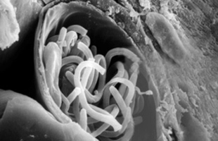
Understanding the pathogenic agent is crucial to understanding any disease; Echinococcosis is no exception. Echinococcosis’s impact as an infectious disease is centered around the actual helminths that cause the disease. And because there are two main forms of Echinococcosis with two different agents — Alveolar and Cystic Echinococcosis — the study of the agent becomes even more crucial.

The global study of Echinococcosis creates a database of knowledge that allows for researchers to understand how the disease varies across the world and what this means for future treatment and prevention. As Rojas, Romig and Lightowlers detail in their 2013 International Journal For Parasitology article entitled “Echinococcus granulosus sensu lato genotypes infecting humans – review of current knowledge,” the different forms of Echinococcosis pathogens call for different responses and treatments. They review the global understanding of the variance in the pathogen, and attempt to discern what this knowledge means for Echinococcosis treatment and prevention going forward.

Cystic Echinococcosis (CE), or hydatid disease, is caused by the larval form of the Echinococcus granulosus tapeworm. As a neglected tropical disease, CE largely affects poorer rural areas with large populations of dogs and sheep. However, not all forms of CE are identical. Through looking at nuclear DNA to understand rNA-encoding regions, different researchers across the world were able to divide the Echinococcus granulosus species into smaller groups (Rojas, Romig and Lightowlers, 2013). Researchers continued to analyze E. granulosus genes using mitochondrial genes, with the most important work differentiating the different species of E. granulosus coming from the work of Josephine Bowles using the cox1 and nad1 genes (Josephine Bowles et al., 1992).

The pathogen can be further classified into ten further genotypes, each genetically unique and given a label (G1-10). Rojas, Romig and Lightowlers (2013) break up the ten genotypes into three main four main groups: Echinococcus granulosus sensu stricto, Echinococcus equinus, Echinococcus ortleppi, and Echinococcus canadensis. Each group has myriad characteristics and differences, from their hosts to the location of the specific form of CE they cause.

G1-3: Echinococcus granulosus sensu stricto

Consisting of three different genotypes, the Echinococcus granulosus sensu stricto genotypic cluster contains far and away the most impactful forms of CE. The G1 genotype alone is responsible for 88.44% of human infections worldwide, and is distributed around the world. Studying the geographical differences among these different genotypes has led to hypothesizing that the Echinococcus granulosus sensu stricto originates in the Middle East and has subsequently spread to other areas. Within its wide range of infective areas — it is present in every continent but Antarctica — certain pockets with high CE prevalence exist where G1 is the only cause of human CE. Its geographical ubiquity can be attributed to the ubiquity of the intermediate hosts associated with Echinococcus granulosus sensu stricto: namely sheep, cattle and water buffalo. These genotypes are also often associated with cosmopolitan distribution, travelling large distances and spanning larger areas.

Echinococcosis Granulosus Genotypes

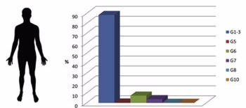
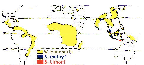
As shown in the chart, genotypes G1-3 (blue) chiefly contribute to human infection.

The important note about the Echinococcus granulosus sensu stricto genotypes is the presence of a vaccine. While these diseases continue to affect the large majority of victims of CE, there is a potential solution. The EG95 protective vaccine antigen has been developed to treat the G1 form of CE specifically. Distributing this vaccine is another matter entirely, but through examining the other genotypes researchers hope to determine whether the EG95 vaccine can protect against multiple forms of CE.

G4: Echinococcus equinus

Only a single genotype, the Echinococcus equinus helminths are most closely associated with its presence in species of the Equidae taxonomic family. Although closely related to the other genotypes of Echinococcus granulosus, Echinococcus equinus has actually never been known to infect humans with CE. This distinction has since been noted in its taxonomic name, as the genotype was initially referred to as Echinococcus granulosus equinus but has since become Echinococcus equinus. It is, however, a specific parasite of Equidae such as horses, donkeys and zebras; Echinococcus equinus is a horse-adapted species that is still present across the world. Recently this G4 genotype was found for the first time in a primate, as Boufana et al. (2012). describe Echinococcus equinus being found in a lemur.

G5: Echinococcus ortleppi

Again a subdivision of E. granulosus consisting of only a single genotype, Echinococcus ortleppi are also not particularly responsible for large-scale human infection of CE. Infections by the G5 genotype have been dubbed “rare” but their similarities to other CE-causing pathogens cannot be overlooked. Echinococcus ortleppi are the adult worm forms of the cattle-associated E. granulosus, and originally adapted to cattle as its intermediate hosts. While the helminth is now extinct in central Europe (its supposed place of origin) it has found hosts in animals across the world. Pigs in Africa, India and South America all carry the G5 genotype. Of more concern to human populations is the recent, albeit slight, rise of G5 in Vietnamese monkeys.

G6-10: Echinococcus canadensis

The largest genotypic cluster of E. granulosus, genotypes G6-10 are all referred to as Echinococcus canadensis. Within this cluster, the existence of the G9 genotype has yet to be fully validated and the G8 and G10 genotypes have only caused cases of Cystic Echinococcosis very rarely. However the G6 and G7 genotypes pose a significantly larger problem for human CE infection. Combined they are responsible for 11.07% of human infections worldwide — with G6 constituting 7.34% and G7 the other 3.73%. The diversity of the Echinococcus canadensis cluster poses challenges for its study and treatment. While only four main genotypes are identified, numerous variants and intermediate forms of Echinococcus canadensis exist. For instance, the G6 genotype exists mainly in Africa and Asia where it is transmitted by camels and goats, and in South America where it is transmitted only by goats; the G7 genotype is mainly existent in Eastern European pigs.


Takeaways & Conclusion

While Rojas, Romig and Lightowlers call attention to the specifics of the varying genotypes of Echinococcus granulosus in “Echinococcus granulosus sensu lato genotypes infecting humans – review of current knowledge” (2013), they end the review with a brief summation of how the different genotypes will influence the future of CE research. The EG95 protective vaccine is their main hope for the prevention of CE, and they determine the vaccine should be trialled to determine its effectiveness for the G6 and G7 genotypes (as well as the G1 genotype it was designed to treat). The variance of the different forms of Echinococcus granulosus contributes to Echinococcosis’s status as a neglected tropical disease, and a furthered understanding of the different agents will lead to advancements and more significant treatments of the disease in the areas that need them most.


The entire article can be found here.

Rojas, Cristian A. Alvarez, Thomas Romig, and Marshall W. Lightowlers. “Echinococcus granulosus sensu lato genotypes infecting humans–review of current knowledge.” International Journal for Parasitology 44.1 (2014): 9-18.

Wuchereria Bancrofti

     Wuchereria Bancrofti are a species of parasitic nematode from the filarias family. Although there are three types of filarias that cause Lymphatic Filariasis, the Wuchereria bancrofti account for around ninety percent of all cases. The bancrofti effects over 100 million people in seventy-six countries within Africa, Central and South America, as well as tropical regions of Asia, Southern China, and the Pacific. The parasites tend to inhabit fresh, still bodies of water in tropical areas, or in swamps or marshes; similar to the mosquito which oftentimes ingests the bancrofti from infected humans, and then pass the larvae to other human hosts.

Screen Shot 2017-03-22 at 10.31.38 PM.png

   The agent starts its life as an egg as forty by twenty-five micrometers, and quickly grows to become 290 by six or seven micrometers in length as a microfilariae, or miniature version of its adult self. In its third stage of development, it is now an infectious larvae, and it is around this time where it develops its notable physical structures like the round mouth with surrounding papillae. After this stage, it is now considered a full adult. A typical adult will be translucent but cloudy with a long, skinny body. Often the tail ends of the nematode will coil or curl up. Females can be around thirty to a hundred millimeters in length while males are typically half the size.

Screen Shot 2017-03-22 at 10.35.21 PM

     The development of the nematodes varies in location. While the parasites are in a human host, they may lay eggs, which then develop into microfilariae, which migrate to blood and lymph vessels of humans. Because nematodes have no circular muscles, the parasites are forced to contract the longitudinal muscles down each of side of its body to move in a forward direction. When a mosquito penetrate the human host, the microfilariae are ingested. Once ingested, they will shed their sheaths, or protective, outer-layer and migrate to the midgut and thoracic muscles of the mosquitoes to develop further. They then use the mosquito as protection as they develop into infectious larvae and migrate to the head of the mosquito so that they are able to be “dropped off” on the next mosquito’s meal from a human host. After initial infection, it takes three to five months to develop into a full-fledged adult. The parasites live for around five years, but can even live up to ten years in some cases.

Screen Shot 2017-03-22 at 10.37.09 PM.png

The Three Species of Mosquitoes which can Host the Bancrofti


     Surprisingly, the Wuchereria bancrofti species requires two parasites of the opposite sex to reproduce. Structurally, the male worms have a corkscrew or coiled region on their tails which allow them to grasp female worms for reproduction. Also, the male genitalia, a “cloaca,” is surrounded with sensilla and spicules which are used to typically to help keep the female nematodes genitalia open for easy sperm transfer. The copulatory spicules act as reference, or guides for the male to physically open the female’s genitalia, while the sensilla, give the male a directional sense as to where it needs to approach, hence the prefix, “sens(e).” There has been evidence that, like many other mating species, the nematodes release chemical pheromones, or exhibit behavior to attract certain females to mate with. While in human hosts, the nematodes live together, coiled up as couples, male and female. Both male and female bancrofti are at sexual and reproductive maturity at around three to twelve months.

Screen Shot 2017-03-22 at 10.38.40 PM.png

     Lacking the ability to hear and see, the bancrofti relies solely on chemosensation to detect chemicals released by other parasites or by the human host. Also, the worms contain surrounding papillae around their mouths and bodies to help strengthen interactions with foods, other nematodes, and again the human environment. The worms use a diurnal rhythm, as well as their chemosensation to match the human lifestyle, by recognizing oxygen blood level to detect whether they are in a vein or arterial vessel compared to the typical lymph vessel. Impressively, this allows them to determine whether or not the human host is sleeping by analyzing blood oxygen levels, to indicate if they should rise to the surface of the peripheral circulation system to be eaten by mosquitoes for the further infection of another host.

While living in human hosts the parasites feed on bodily fluids and tissue. The bancrofti lodge themselves in lymph vessels which causes massive swelling, but their residency allows them to be surrounded by food at all times. Luckily for them, there are no known predators which can prey upon them while in the human host. Humans are in fact the only known natural host for the bancrofti, but there have been successful attempts to infect species of monkeys with the parasite. The bancrofti can live in four different hosts; one if which is a human, and the other three are all mosquito species.

Onchocerciasis


Onchocerciasis-River Blindness

Agent – What is the causative agent? Scientific name? Image? Type of pathogen? Where is it found?

Onchocerca volvulus is the pathogen responsible for the disease onchocerciasis, or river blindness. Onchocerciasis is caused by nematodes (roundworms) that inhabit subcutaneous tissues. Onchocerciasis affects at least 17.7 million people worldwide. It is  a non-fatal but severely debilitating disease. It has been found in 37 countries (30 in Africa, 6 in the Americas, and Yemen).

Onchocerciasis is transmitted through the bites of Simulium blackflies. Blackflies breed in fast-flowing rivers and streams, increasing the risk of infection to people living nearby.Adult Onchocerca volvulus worms can live for fifteen years in the human body.  Common blackfly hosts include Similium damnosum and S. neavei in Africa and S. ochraceum, S. metallicum, S. callidum, and S. exiguum in the Americas.

adult_wormsonchocerca_volvulus-adult-worms


Host – How are humans affected? Which systems are impacted? What treatments are available?

Onchocerciasis occurs when a blackfly bites an infected person during a blood meal. During the bite, microfilariae of the Onchocerca volvulus  are transferred to the person from the fly. The microfilariae then develop into infected larvae over the next few weeks. Live microfilariae are barely noticed inside the body as they cause little inflammation, but dead and degenerating microfilariae in the skin cause severe dermatitis, intense itching, skin depigmentation, skin thickening, skin discoloration, cracking of skin, and loss of skin elasticity.

Quite often, microfilariae migrate to the eye, and their death causes intense inflammation. Over years this sclerosing (scarring) keratitis hardens the cornea and causes blindness.Adults can be found knotted together in pairs or groups in the subcutaneous tissues.

Treatments

There are a few treatment options for onchocerciasis. One is a surgical operation to remove onchocercomas (especially those around the head) in order to reduce the rate of visual impairment and the number of new infections.

The most popular treatment for infected patients is chemotherapy with the drug ivermectin. A single oral dose given annually eliminates skin microfilariae, interrupts the transmission cycle by suppressing adult female microfilariae release, improves skin disease, and eventually kills adult worms. need to be given every 6 months for the life span of the adult worms or for as long as the infected person has evidence of skin or eye infection. Ivermectin kills the larvae and prevents them from causing damage but it does not kill the adults. There is a promising new treatment using doxycycline that kills the adult worms. They do this by killing the Wolbachia bacteria on which the adult worms depend in order to survive.

Thirdly, larvicides have been used to reduce the blackfly vector population. A process to spay chemicals over high concentrations of black have proved very successful. Between 1974 and 2002 using mainly the spray of insecticides against black-fly larvae (vector control) by helicopters and airplanes and upplemented by large-scale distribution of ivermectin since 1989, the OCP (Onchocerciasis Control Program) relieved 40 million people from infection. These measures prevented blindness in 600 000 people, and ensured that 18 million children were born free from the threat of the disease and blindness.


Environment – What environmental or behavioral factors result in disease transmission? What environmental or behavioral factors impact treating the disease.

Blackflies breed in fast-flowing rivers and streams, increasing the risk of infection to people living nearby. Transmission of the disease is through the bite of female blackflies (usually occurring during the daytime near rapidly flowing rivers and streams). Multiple bites are usually needed before being infected.

There are no vaccines or medications available to prevent becoming infected with O. volvulus. The best prevention efforts include personal protection measures against biting insects. This includes wearing insect repellant such as N,N-Diethyl-meta-toluamide (DEET) on exposed skin, wearing long sleeves and long pants during the day when black flies bite, and wearing permethrin- treated clothing.

http://animaldiversity.org/accounts/Onchocerca_volvulus/#economic_importance_positive

https://www.cdc.gov/parasites/onchocerciasis/biology.html

http://www.who.int/onchocerciasis/epidemiology/en/

http://www.who.int/apoc/onchocerciasis/lifecycle/en/

http://www.medicinenet.com/onchocerciasis/article.htm